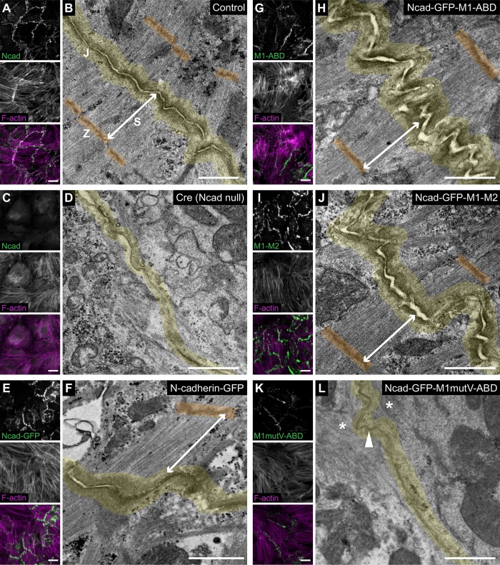
FIGURE 6:
Vinculin recruitment is required to couple myofibrils to the AJ. Ncadfx/fx cardiomyocytes uninfected (A, B), infected with Cre (C, D), infected with Cre and Ncad-GFP (E, F), or infected with Cre and N-cadherin-GFP-αE-catenin fusion adenoviruses (G–L) were fixed and processed for immunostaining or thin-section TEM. (A, C, E, G, I, K) Immunofluorescence lower-magnification (40×) images of control and infected cardiomyocytes. Uninfected control (A) and Cre-infected (C) cardiomyocytes were stained for N-cadherin and F-actin. Ncad-GFP (E) and N-cadherin-GFP-αE-catenin fusion adenovirus-infected (G, I, K) cardiomyocytes were stained for F-actin. Individual and merged N-cadherin/GFP (green) and F-actin (magenta) channels shown. Images are maximum projections of 5 μm stacks. (B, D, F, H, J, L) Representative TEM image of a cell–cell contact from >60 images from at least three independent experiments. Cell–cell junctions (J) are highlighted in yellow, Z-discs (Z) are colored orange, and a terminal myofibril sarcomere (S) is defined by a double arrow line. In L, the arrowhead marks electron density along the cell–cell junction and the asterisks mark poorly organized F-actin. Scale bar is 20 µm in A, C, E, G, I, and K and 1 μm in B, D, F, H, J, and L.

