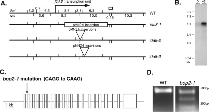
FIGURE 1:
Molecular characterization of ida8 and bop2 mutations. (A) Diagram of the ∼40 kb region of genomic DNA around the IDA8 locus in WT, with SacI restriction sites indicated on top and Not1 restriction sites indicated below. The location of the IDA8 transcription unit is shown by the arrow. The site of the genomic fragment recovered by plasmid rescue from ida8-1 is shown by the white box. The next three lines show the sites of pMN24 insertion in each ida8 allele as determined by Southern blotting (Supplemental Figure S1E). (B) Northern blot of total RNA isolated from WT cells before (0) and 45 min after deflagellation and probed with a 6.5 kb SacI restriction fragment that was missing in ida8-1. Other blots probed with the 5.6 and 2.3 kb SacI fragments and several RT-PCR products recognized the same transcript. (C) Diagram of the intron-exon structure of the IDA8 gene showing the bop2-1 mutation in the acceptor splice site of the second exon. (D) RT-PCR products obtained from WT and bop2-1 RNA using primers surrounding the site of the bop2-1 mutation were analyzed on an agarose gel. Sequence analysis identified premature stop codons in all of the RT-PCR products from bop2-1.

