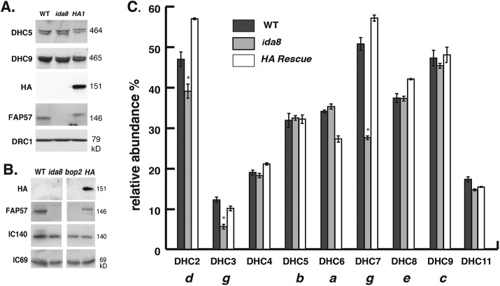
FIGURE 5:
Mass spectrometry reveals defects in the assembly of a subset of inner arm DHCs in ida8. (A) Western blot of axonemes from WT, ida8-1, and an ida8-1; FAP57-HA rescued strain (HA1) was probed with antibodies against two inner arm DHCs (DHC5, DHC9), HA, FAP57, and DRC1. (B) Western blot of axonemes from WT, ida8-1, bop2, and bop2; FAP57-HA was probed with antibodies against HA, FAP57, IC140, and IC69. (C) The axoneme samples shown in A were fractionated by SDS–PAGE, and the DHC region was excised and analyzed in triplicate by tandem MS/MS and spectral counting. The total counts for each DHC were expressed as a percentage of the total counts for the two I1 dynein DHCs. The bars represent the range of the three replicates, and the asterisks represent those DHCs that were reduced more than 15% in ida8-1 relative to both the WT and the HA1 rescued strains.

