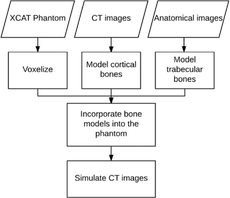
Fig. 1.
Flowchart of the bone modeling. XCAT phantoms were used as the basis for each model. Cortical bones were modeled based on cortical thicknesses of corresponding CT images of each XCAT. Trabecular bones were modeled using high resolution anatomical images. Finally, each phantom’s realism was objectively evaluated by simulating its CT images.

