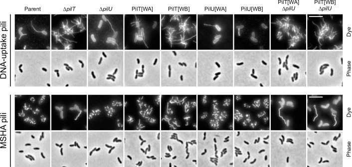
Fig 3. Cysteine labelling of DNA-uptake and MSHA pili in PilT and PilU variants.
Visualisation of DNA-uptake pili (PilA[S67C]) and MSHA pili (MshA[T70C]) in the indicated backgrounds. To visualise DNA-uptake pili strains were cultured in the presence of chitin-independent competence induction. Cells were stained with AF-488-Mal (Dye). Scale bars = 5 μm.

