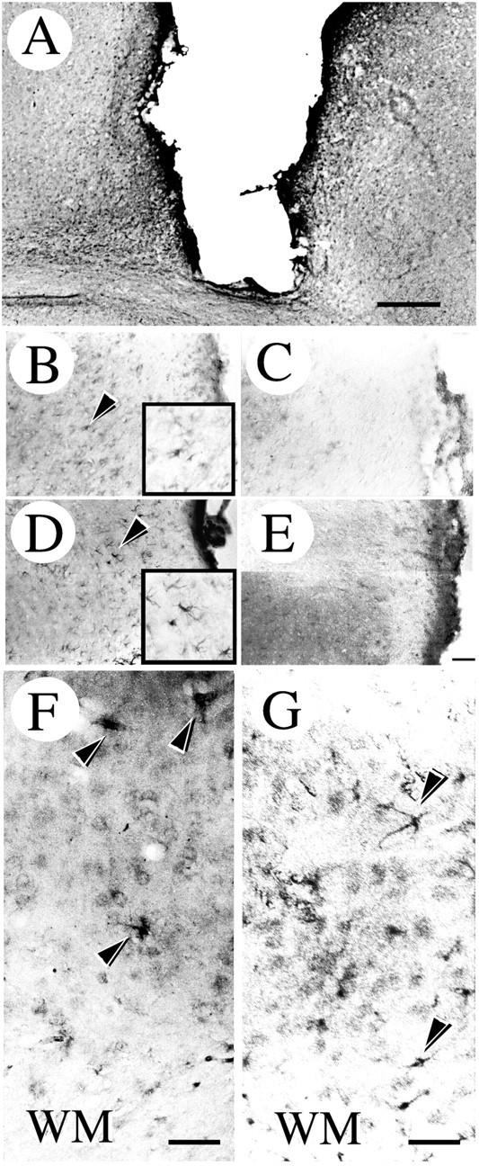
Fig. 3.

Immunohistochemical localization of ErbB2 and ErbB3 in the injured brain. A shows anti-ErbB2 immunoreactive cells appearing near the wound. B, D, Higher magnification images of an area adjacent to the wound stained with anti-ErbB2 and anti-ErbB3 antibodies, respectively. The immunopositive cells indicated by the arrowheads are shown at a higher magnification in the box. These immunopositive cells were not observed in the sections stained with antibodies preabsorbed with the corresponding recombinant peptides (C, E). In the deep layer of the injured cortex, ErbB2 (F) and ErbB3 (G) immunoreactivities were also detected. The black arrowheads shown in F and Gindicate reactive astrocyte-like cells. WM, White matter. Scale bars: A, 0.5 mm; B–E(shown in E), 50 μm; F, G, 20 μm.
