
Fig. 5.
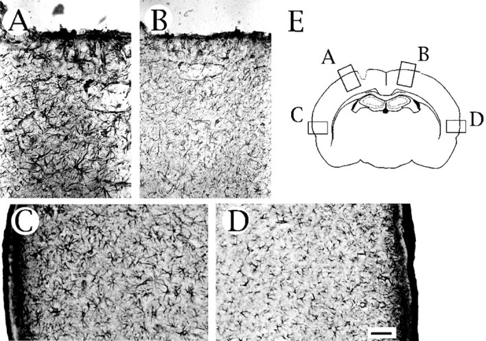
Immunohistochemical localization of an astrocytic differentiation marker, GFAP, in the injured brain. Intense immunoreactivity for GFAP is observed in the vicinity ofA and even in an area far from the injured site in the ipsilateral hemisphere (C), but lower GFAP immunoreactivity was observed in the contralateral hemisphere (B, D). E, Schematic representation of the areas used for the microscopic observation. Scale bar (in D), 50 μm.