
Fig. 8.
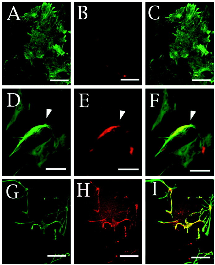
Immunofluorescence staining of neuregulin and GFAP on cultured astrocytes. Astrocytes cultured in DMEM supplemented with 10% FCS in the absence (A–F) or presence (G–I) of 3 μm forskolin for 3 hr are shown. The polygonal flat cells (A–F) and the well characterized morphologically differentiated cells induced by forskolin administration (G–I) were double-stained with anti-GFAP (A, D, G) and anti-neuregulin (B, E, H) antibodies. Anti-neuregulin and anti-GFAP antibodies were visualized with a biotinylated anti-goat IgG antibody and Cy3-labeled streptavidin (red) and fluorescein-labeled anti-rabbit (green) IgG antibodies, respectively. Thewhite arrowhead in D–E indicates a neuregulin-positive cell in the control culture. The merged images of A and B, D andE, and G and H are also shown in C, F, and I, respectively. Scale bars, 10 μm.