
Fig. 4.
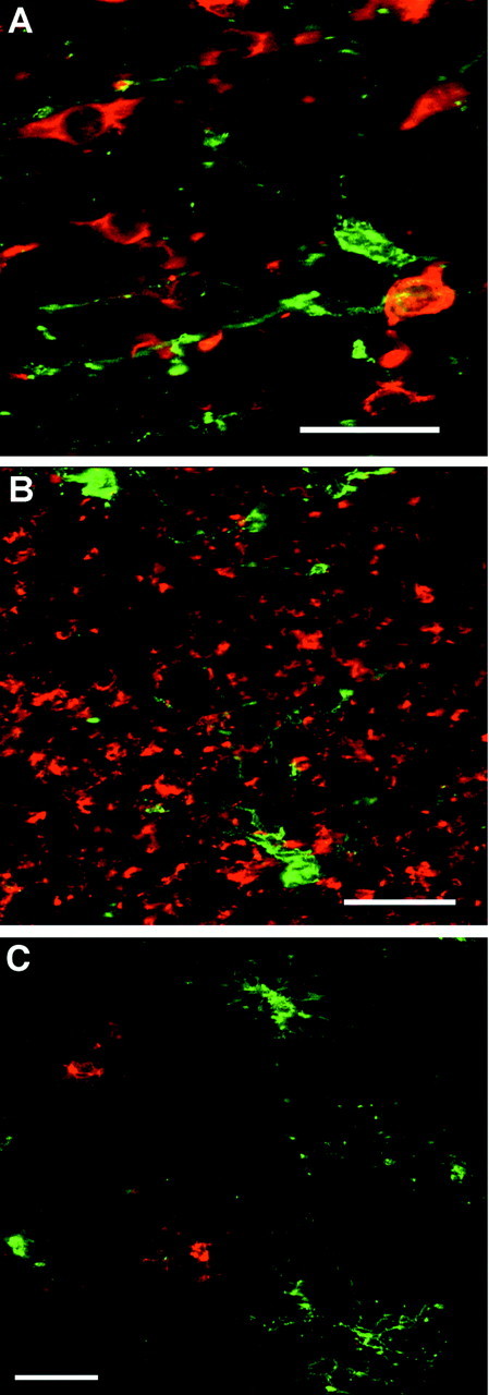
Representative confocal laser microscopic digital images demonstrate the specificity of the O4 antibody for OL precursors. A, B, O4-labeled cells (green) differ in morphology and distribution from neuronal somata (red) in the cortical mantle (A) and from neuronal processes (red) in the superficial white matter (B) that were visualized with anti-β-tubulin isotype III. C, Microglia labeled with the R. communis lectin (red) have a similar morphology but a distinctly different distribution from that of O4-labeled cells (green) in the superficial white matter. Scale bars: A, 20 μm; B, C, 25 μm.