
Fig. 5.
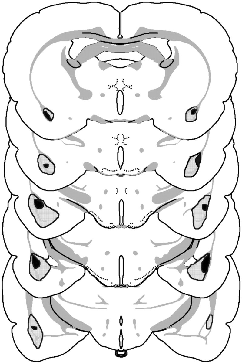
Schematic representation of excitotoxic lesions to the basolateral amygdala from experiment 2. Shaded areasrepresent the smallest (black) and largest (gray) extent of neuronal damage. Coronal sections are −1.8 to −3.8 mm relative to bregma (Swanson, 1998).