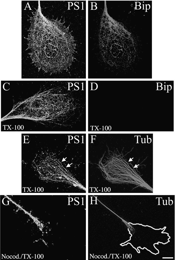
Fig. 1.
PS1 associates with MT in neuronal growth cones. Confocal images show the distribution of PS1 (A) and Bip (B) in 3 DIV hippocampal growth cones. PS1 colocalizes with Bip at the central region of the growth cone. Note that PS1 IF extends to filopodia and lamellipodia (A). Cultures were stained sequentially with PSN2 and anti-Bip antibodies (see Materials and Methods). After TX-100 extraction, PS1 IF remains in the cytoskeletal preparation (C), whereas Bip IF is abolished completely (D). Shown is double IF for PS1 with antibody 2025 (E) and β-tubulin class III (F). PS1 IF appears closely associated with the microtubular network. Partial colocalization between PS1 and individual MT can be observed (arrows, E, F). A mild nocodazole treatment (see Materials and Methods) abolished PS1 (G) and β-tubulin class III (H) IF from neuronal growth cones of TX-100-extracted cultures. Note the presence of PS1 IF in the neuritic shaft where intact MT are still present (G, H). The perimeter of the growth cone before nocodazole treatment is shown in H. Scale bar, 10 μm.

