Fig. 7.

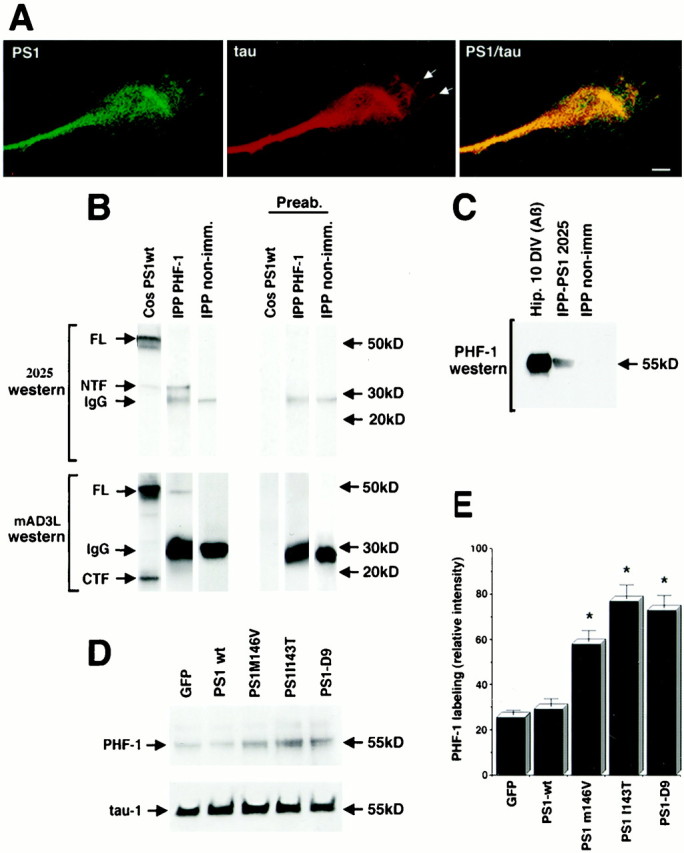
PS1 colocalizes and coimmunoprecipitates with tau, and PS1 mutations increase Aβ-induced tau phosphorylation.A, Double IF showing colocalization of endogenous PS1 (PSN2) and tau (Tau-1) in neuronal growth cones. Note the presence of tau IF in filopodia (arrows). Scale bar, 10 μm.B, Cell lysates of 10 DIV hippocampal neurons treated with Aβ were immunoprecipitated with PHF-1 (IPP PHF-1), blotted, and stained with antibodies 2025 and mAD3L. Antibody 2025 recognized PS1 NTF in PHF-1-immunoprecipitated material. Antibody 2025 also labeled PS1 FL and NTF in a homogenate of transfected Cos cells (Cos PS1wt) used as a positive control. Antibody mAD3L labeled PS1 FL and CTF in a homogenate of transfected Cos cells and recognized a band corresponding to PS1 FL in PHF-1-immunoprecipitated material. Cell lysate immunoprecipitated with nonimmune mouse sera (IPP non-imm) showed no reaction with 2025 and mAD3L antibodies. Preabsorption of both 2025 and mAD3L with the corresponding antigenic peptides completely abolished PS1-specific labeling in the Western blot (Preab). Secondary antibodies used to develop the immunoblots reacted with PHF-1 IgG in the immunoprecipitated material (IgG).C, Cell lysates of 10 DIV hippocampal neurons treated with Aβ were immunoprecipitated with anti-PS1 antibody 2025, blotted, and reacted with PHF-1, which recognizes phosphorylated tau at Ser 396/404. PHF-1 labeled a 55 kDa band in a homogenate of Aβ-treated neurons (Aβ) and a band of similar molecular weight in the material immunoprecipitated with 2025 (IPP-PS1 2025). Cell lysate immunoprecipitated with nonimmune rabbit sera (IPP non-imm) showed no reaction with PHF-1.D, Western blot analysis of transfected cultures treated with Aβ. Cultures transfected at 8 DIV were treated with 20 μm Aβ and harvested at 10 DIV. Protein (10 μg) was loaded in each lane. The blots were developed with PHF-1 and Tau-1 antibodies. Expression of green fluorescent protein (GFP) was used as a control. Note the increase in PHF-1 staining in cultures expressing PS1 mutations. No significant changes in the levels of nonphosphorylated tau were detected by Tau-1. E, Relative changes in the level of phosphorylated tau in Aβ-treated cultures expressing PS1 mutations detected by the PHF-1 antibody. Quantitative Western blot analysis was performed as described in Materials and Methods. Values are the mean ± SE; n = 5 independent experiments. *p < 0.01 relative to PS1-wt by Student's t test.
