
Fig. 7.
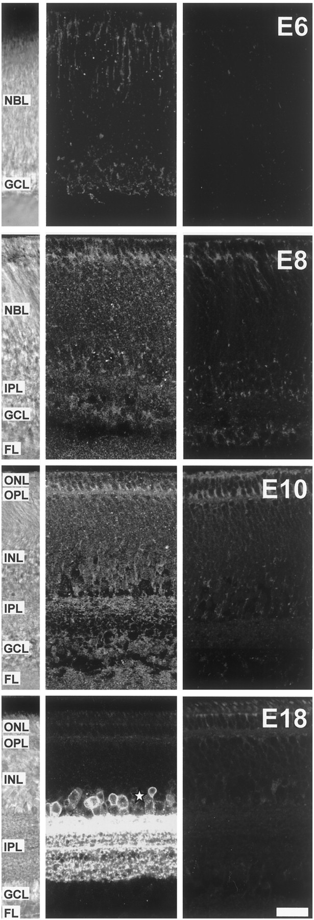
Expression of GAT-1 transporters in the developing chick retina. Confocal microscope sections of GAT-1 transporter immunostaining of transverse vibratome slices from E6, E8, E10, and E18 chick retina. Left panels, White-light micrographs from the slices in the middle panels showing the structure of the retina for each age. Middle panels, Sections immunoreacted with the anti-GAT-1 antibody. Right panels, Negative controls in which the primary antibody was omitted. At E6, GAT-1 immunostaining was punctate and restricted to the outer NBL and the GCL. At E8, immunoreactive puncta were distributed throughout the retina. By E10, GAT-1 lamina-specific staining was apparent in the IPL, with laminas closer to the INL more strongly labeled. At E18, GAT-1 staining is restricted to the IPL, in which it has a clear laminar distribution, and to somata in the proximal INL, presumptive amacrine cells (asterisk). Some autofluorescence is apparent in the outer retina at the level of the ONL and OPL (see negative controls). Scale bar, 20 μm.