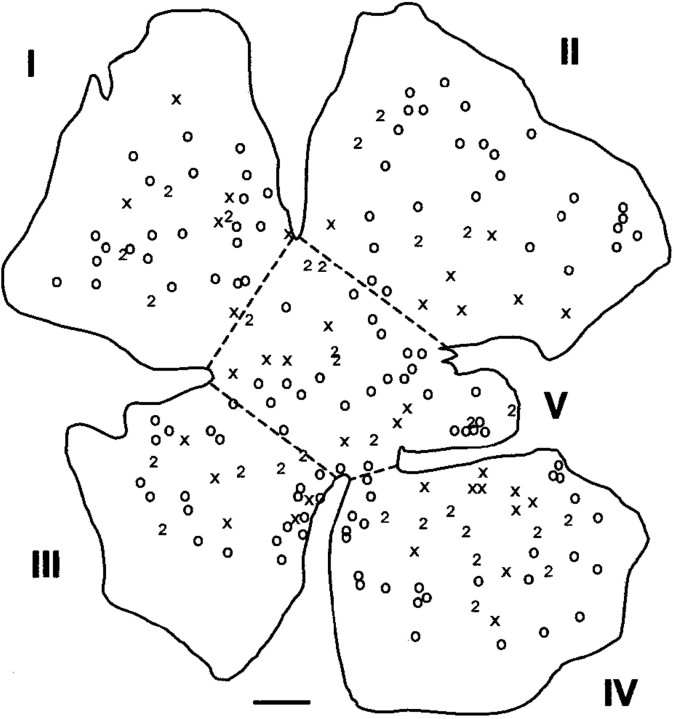
Fig. 4.
Camera lucida drawing of retrogradely labeled RGCs in a whole mount of a retina from which RGC axons had regenerated into a peripheral nerve graft. The graft was divided into two branches labeled with fast blue (crosses) or fluorogold (open circles). Double-labeled cells are indicated by the number 2. The whole mounts prepared in this manner were arbitrarily divided into five approximately equal sectors (I–V) as illustrated to permit evaluation of the proportions of labeled cells in various areas of the retina. Scale bar, 500 μm.

