
Fig. 1.
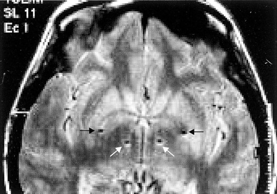
T1-weighted magnetic resonance axial view in a patient with Parkinson's disease after implantation of the left then right pallidum and STN. The sites of the deepest contacts (0) are shown and lie in STN (white arrows) and GPi (black arrows), bilaterally. The scale to the right is in centimeters.