
Fig. 6.
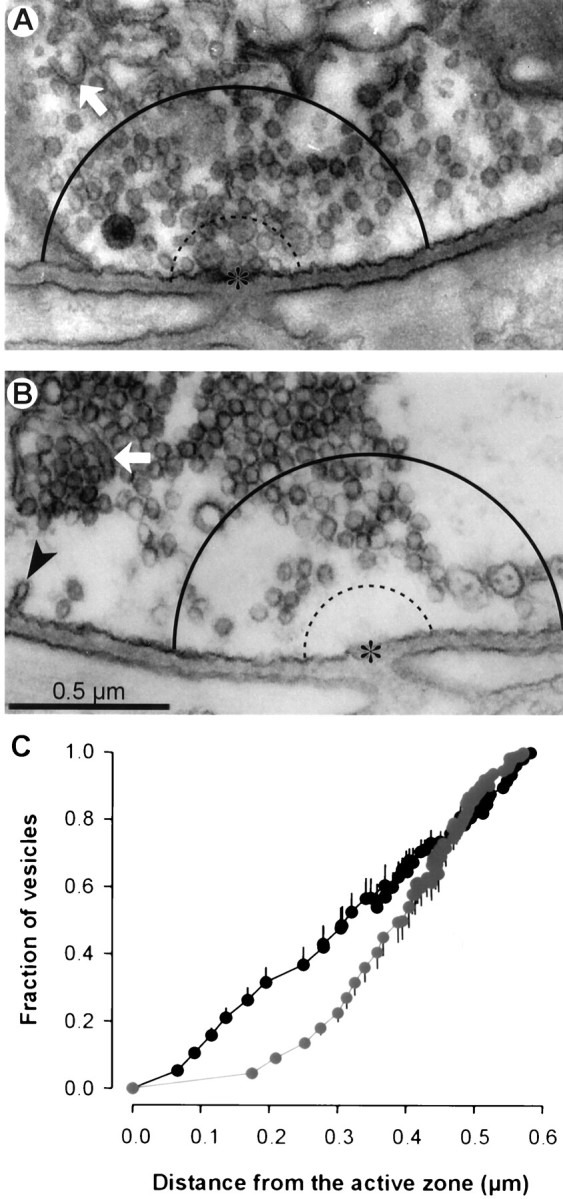
Staurosporine caused vesicle depletion near active zones after tetanic stimulation. Preparations were fixed immediately after stimulation (30 Hz for 2.5 min) and prepared for electron microscopy. Active zones (asterisks) were identified by adjacency to openings of postsynaptic folds. Vesicles in control (A) and staurosporine-treated (B) terminals lying within 0.6 μm (solid curved line; dotted line shows 0.2 μm distance) were counted, and distances to the nearest active zone were measured. Cisternae (arrow) and coated pits (arrowhead) often were observed. C, The average fraction of vesicles (n = 16 terminals) located within a given distance from the active zone reveals a depletion of vesicles near active zones in staurosporine-treated terminals (gray) as compared with controls (black). Error bars show ± SEM.