
Fig. 5.
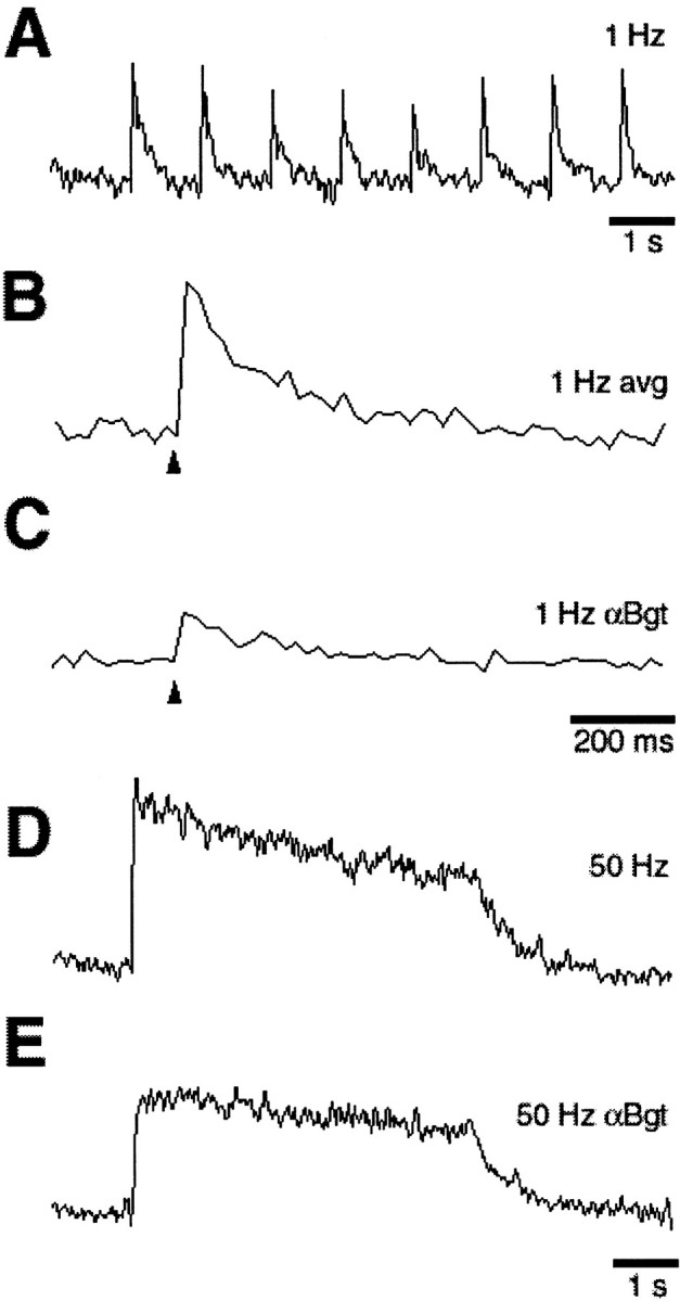
Synaptically driven calcium transients in ciliary neurons from posthatch chicks. Ciliary ganglia from 3-week-old chicks were dissected, loaded with dye, stimulated, and imaged as described for E15 ganglia in Figures 1-4. A–C, On-spine calcium transients elicited by 1 Hz synaptic stimulation and shown either as a continuous trace or as a signal averaged over eight responses either before or after a 30 min exposure to α-Bgt to block α7-nAChRs, as indicated. D, E, On-spine calcium elevations induced by 50 Hz synaptic stimulation before and after α-Bgt treatment. As in E15 ganglia, synaptic stimulation at 1 Hz induced rapid calcium transients on-spine, and the transients were almost completely dependent on α7-nAChRs; 50 Hz stimulation induced a sustained elevation showing a biphasic decay, and α7-nAChR blockade eliminated the rapidly decaying component. Similar results were obtained with all six ganglia that were tested (one neuron per ganglion). Calibration: A, D, E, 1 sec; B, C, 200 msec. Arrowheads indicate the time of stimulation.