
Fig. 7.
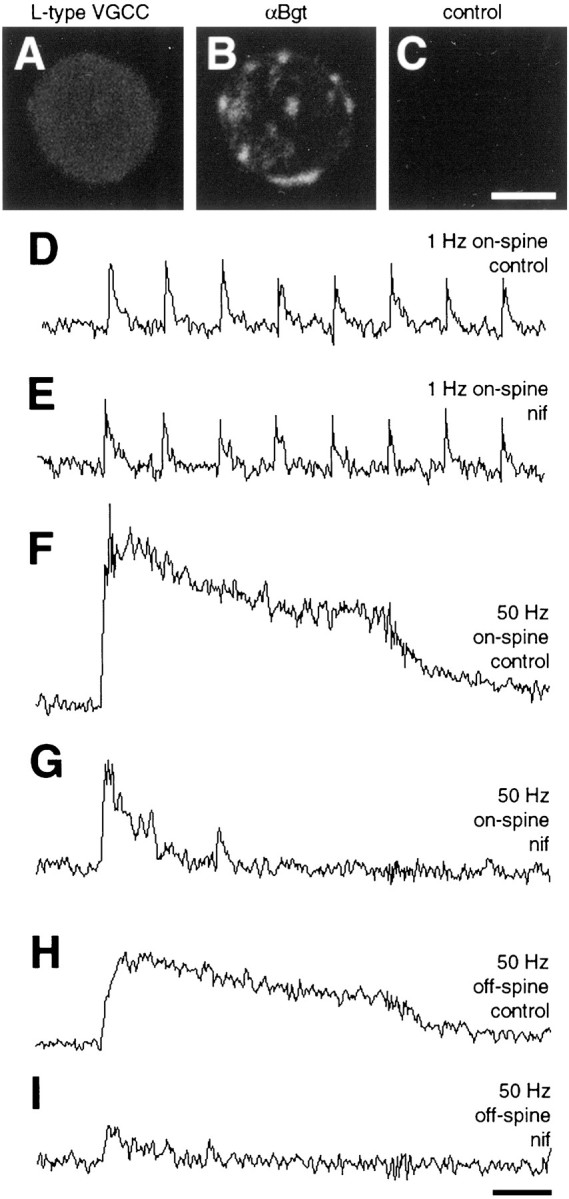
Contribution of voltage-gated L-type calcium channels to synaptically driven calcium elevations. A, Immunofluorescent detection of voltage-gated L-type calcium channels on a dissociated E15 ciliary neuron, using an antibody to the α1c subunit and fluorescent secondary antibody.B, Same neuron as in A costained with rhodamine-α-Bgt to reveal the α7-nAChRs clusters representing somatic spine mats. C, Control showing absence of labeling for an E15 neuron incubated with rabbit IgG instead of the α1c antibody. The same gain and exposure times were used for A–C. D–I, Continuous calcium imaging of on-spine and off-spine regions of a neuron stimulated at 1 or 50 Hz, as indicated, before (control) and after (nif) a 30 min incubation with 10 μm nifedipine to block voltage-gated L-type calcium channels. Almost all of the slowly decaying cell-wide calcium elevation was lost when L-type calcium channels were blocked; much of the fast-decaying on-spine calcium transient remained. Similar results were obtained for all nine neurons that were tested. Scale bar forA–C, 10 μm; calibration for D–G, 1 sec.