
Fig. 2.
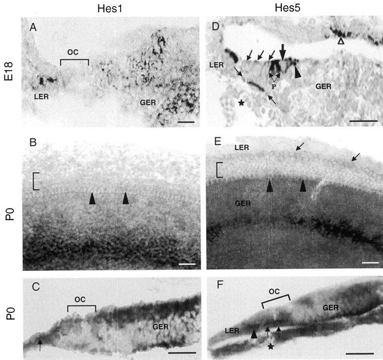
Nonradioactive Hes1(A–C) and Hes5(D–F) RNA in situ hybridization in the developing mouse cochlea at E18 and P0. A,D, In situ hybridization on transverse sections through the base of E18 cochlea. Hes1 mRNA (A) is expressed in the LER cells and in a large area of GER, except in the organ of Corti (OC).Hes5 mRNA (D) is expressed in the LER, pillar cells (P), and Deiter's cells at the base of OHCs (area bracketed by arrows).Hes5 is also found in the inner phalengeal cells (arrowhead), which are adjacent to the inner hair cell region. Hair cells (large arrow indicates IHC;oblique arrows indicate the three rows of OHCs) are lacking Hes5 hybridization signal. Note theHes5 expression in the marginal cells of the stria vascularis (open arrowhead). Asteriskindicates the spiral vessel, a landmark for the location of the organ of Corti at this stage of maturation. B,E, In situ hybridization on whole-mount surface preparations from the middle turn of the cochlear duct at P0. Expression of Hes1 (B) spans a large area of the GER, as observed in a cochlea section (A). Expression of Hes5(E) is observed in a relatively narrow band of cells of the GER. There is a faint but specific Hes5expression in the cells within the LER (arrows). Note the absence of both Hes1 and Hes5hybridization signal from the hair cells (arrowheadsindicate the location of IHC row; bracket indicates the OHC rows). C, F, Transverse sections through the whole-mount cochlear surface preparations presented inB and E, respectively. C, This section confirms Hes1 expression in the GER cells and its lack from the cells of the organ of Corti (bracket). Hybridization label for Hes1is also located in the LER (arrow). F,Hes5 expression is restricted to the nonsensory supporting cells (Deiter's cells; arrows) within the organ of Corti. This section also confirms the specific expression ofHes5 is the LER cells (arrowhead). A weak label for Hes5 is observed in the GER region and in cells located near the basilar membrane. Asteriskindicates the spiral vessel. Scale bars, 20 μm.