
Fig. 3.
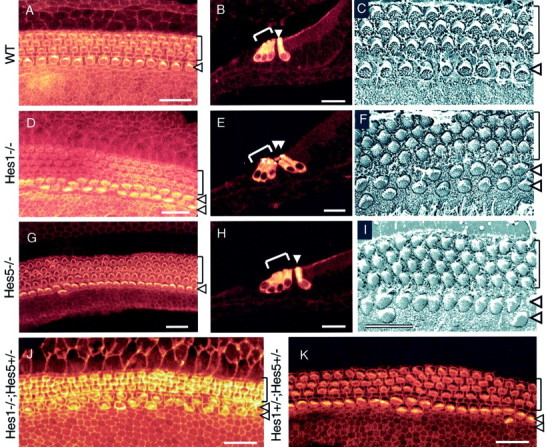
A comparison of hair cell development in P0 cochleae from wild-type control (A–C),Hes1−/−(D–F),Hes5−/−(G–I), andHes1−/−;Hes5+/−(J) mutants andHes1+/−;Hes5+/−heterozygous mice (K). Lack ofHes1 and Hes5 causes the development of supernumerary hair cells. A, D,G, J, K, Confocal images of surface preparations stained with rhodamine phalloidin to visualize the actin-rich stereocilia of the hair cells.B, E, H, Cross-sections through the organ of Corti in the midmodiolar region immunostained with antibody anti-myosin VIIa. C, F,I, Scanning electron microscopy of the surface of the organ of Corti in the midcochlear turn. In control cochlea, the normal pattern is well defined, with a single row of IHCs and three rows of OHCs. In contrast, in Hes1−/−cochleae, two rows of IHCs and three to four rows of OHCs are present. In Hes5−/−cochleae, four rows of OHCs are often present, in addition to dispersed regions along the sensory epithelium that contain few IHC pairs. J,Hes1−/−;Hes5+/−cochleae revealed the same phenotype asHes1−/−, but the effect on the number of supernumerary hair cells was more important inHes1−/−;Hes5+/−cochleae (see Table. 1). K, Cochlear surface preparation from double heterozygous mouse cochlea indicating regions with extra OHC rows and a few pairs of IHCs. Brackets mark the OHCs rows, and arrowheads point to the IHC row. Scale bars: A, B, D,E, G, H, J,K, 20 μm; C, F,I, 10 μm.