
Fig. 7.
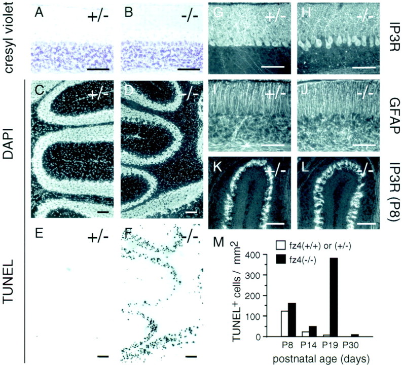
Extensive granule cell death between weeks 2 and 5 in the fz4(−/−) cerebellum.A–J, Cerebella fromfz4(+/−) and fz4(−/−) mice at P19.A, B, Cresyl violet staining reveals a grossly normal appearance including roughly normal numbers of Purkinje and granule cells. DAPI staining (C, D) and TUNEL labeling of DNA fragments (E,F) in the same sections reveal extensive granule cell death in the fz4(−/−) cerebellum (F) and minimal cell death in thefz4(+/−) control (E).G, H, Type I IP3 receptor immunostaining at P19 shows increased separation of Purkinje cell bodies and processes in the fz4(−/−) cerebellum.I, J, Activated glia, as revealed by GFAP immunostaining, show subtle disorganization in thefz4(−/−) granule cell layer. K,L, Type I IP3 receptor immunostaining at P8 shows that developing Purkinje cells of fz4(+/−) andfz4(−/−) mice have indistinguishable morphology.M, Histogram showing, at different ages, the number of TUNEL-positive nuclei per square millimeter of surface area in a series of randomly selected 16-μm-thick sections of cerebellum. Because cell sizes are smaller and cell densities are higher in younger animals, the total number of cells per square millimeter is higher in younger animals. Scale bars, 100 μm.