
Fig. 1.
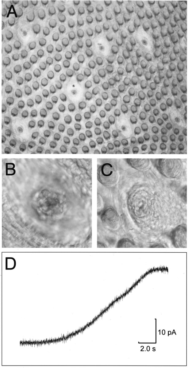
Photomicrographs of the lingual epithelium, after enzymatic removal from the anterior portion of the tongue.A, At low power, viewed from the serosal (basolateral) side of the epithelium, several taste buds within fungiform papillae can be seen amid the numerous filiform papillae in a freshly isolated strip of epithelium. B, At higher power and under DIC illumination, individual cells in the taste bud can be visualized.C, Taste pore of an individual taste bud, viewed from the mucosal side of the epithelium. Tastants applied to the mucosal surface have access to the taste bud only through the taste pore.D, Change in current produced by the liquid junction potential of a micropipette filled with 30 mm KCl when the mucosal solution was switched from 30 to 300 mm KCl.