
Fig. 6.
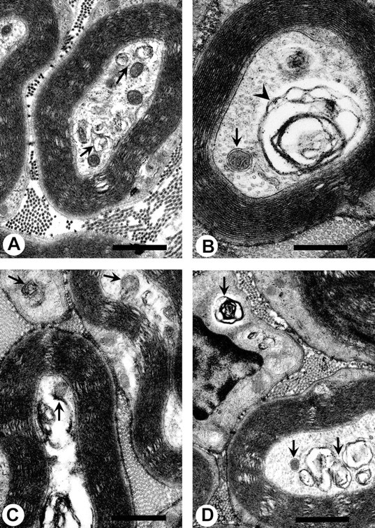
A–D, Pathological lesions in tibial nerves of T mice chronically injected with 2,5-HD (8 mmol · kg−1 · d−1) for 19 d. Typical lesions include accumulations of mitochondria, dense bodies, and other membrane-bound vesicles (arrows), as well as the presence of multilaminar bodies (arrowheads). These lesions are very similar to those observed in NT animals, except neurofilaments are absent from these nerve sections (see Fig. 7 to compare). These lesions are also similar to those observed with chronic ACR exposure (see Figs. 4, 5 to compare). A–D, 21,500×. Scale bars, 1 μm.