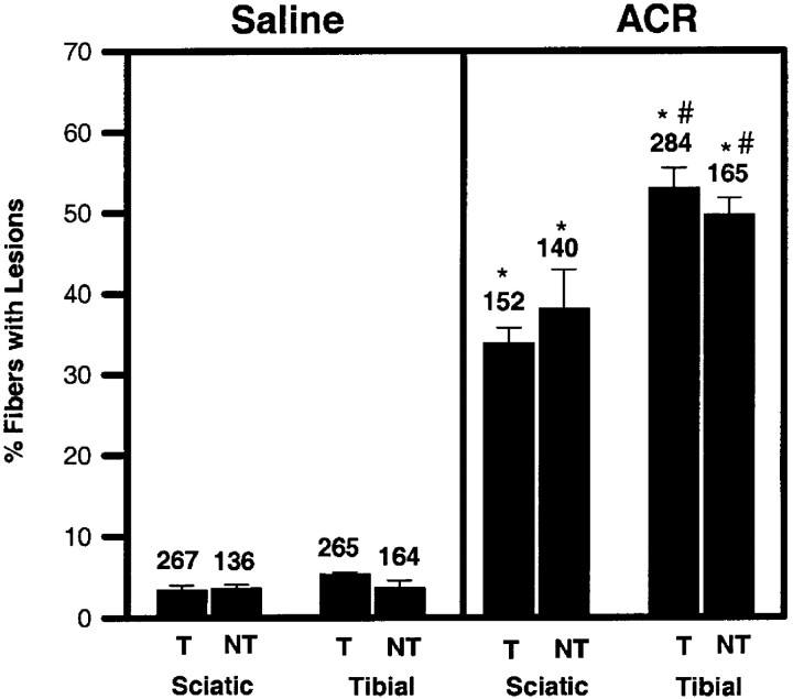
Fig. 8.
Quantitative comparison of frequency of pathological lesions within axons of sciatic and tibial nerves of T and NT mice injected with 50 mg · kg−1 · d−1 ACR or saline for 18 d. The number of axons counted in each group is provided above each bar. ACR significantly increased the frequency of lesions over controls. A significant difference was also observed between sciatic (proximal) and tibial (distal) nerves in both T and NT mice. No differences were found between T and NT mice under any experimental condition. Comparable data for 2,5-HD were unavailable because of an unresolvable difficulty in the perfusion of 2,5-HD-exposed mice. Statistical differences were determined using a two-way ANOVA for repeated measures followed by Tukey's highly significant differences post hoc test. *p < 0.05, significantly different from the corresponding saline control; #p < 0.05, significantly different from the corresponding sciatic nerve.

