Fig. 3.
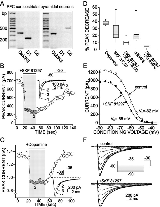
D1/D5 receptor activation reduces the rapidly inactivating Na+current. A, The scRT-PCR revealed that D1and/or D5 mRNA was expressed in PFC neurons. Shown are photographs of gels derived from two pyramidal neurons expressing CaMKII mRNA. One had detectable levels of D1 receptor mRNA; the other had detectable levels of D5 receptor mRNA. The sizing ladder is in the left-most lane of both gels.B, Plot of peak Na+ current evoked by a step from a holding potential of −80 to −30 mV as function of time. D1/D5 receptor agonist SKF 81297 (1 μm) reversibly suppresses the peak current.Inset, Representative currents used to constructB. C, Plot of peak Na+current evoked by a step from a holding potential of −80 to −35 mV as function of time. Dopamine application (50 μm) also reversibly suppresses the peak current. Inset, Representative currents used to construct C.D, Box plot summary of the modulation of Na+ transient current. SCH 23390 (1 μm) blocked the effect of SKF 81297 (1 μm;n = 7). This effect was mimicked by cBIMPS (50 μm; n = 7), a PKA activator, and was blocked by Rp-cAMPS (10 μm; n = 4), a PKA inhibitor, indicating the involvement of the PKA in the response that was observed. E, A representative steady-state inactivation plot derived from a PFC pyramidal neuron. SKF 81297 (1 μm) reduced the maximum current evoked by a step to −30 mV and shifted the voltage dependence of steady-state inactivation to slightly more negative potentials (Control:Vh = −62 mV,Vc = 5 mV; SKF:Vh = −65 mV,Vc = 4.9 mV). F, Current traces and protocols used to construct the steady-state inactivation plot shown in D.

