
Fig. 1.
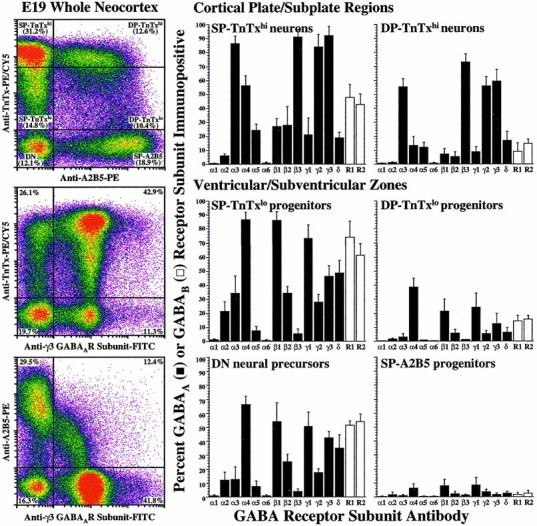
GABAA receptor subunits are differentially expressed during neuronal lineage progression. Using quantitative flow cytometry of TnTx/A2B5 surface-labeling patterns and intensities in conjunction with microdissection, we identified the E19 neocortical cells as neural precursors (DN), neuroglial (SP-A2B5) or neuronal (SP-TnTxlo or DP-TnTxlo) progenitors from the VZ/SVZ, or postmitotic/postmigratory differentiating neurons (SP-TnTxhi or DP-TnTxhi) from the CP/SP regions (Maric et al., 2000b). After surface phenotyping, the cells were fixed and processed for the expression of 13 GABAA receptor and 2 GABAB receptor subunits (see Materials and Methods).Left, As an example, the log–log dot density plots inpseudocolor show the TnTx/A2B5 labeling pattern characteristic of E19 neocortical cells and the distributions of the γ3 GABAA receptor subunit in the context of either surface label. Boundaries (crosshairs) between labeled and unlabeled cells and between cells expressing low and high levels of TnTx labeling (TnTxlo and TnTxhi) have been drawn empirically to quantify the percentages of cells in each subpopulation. Middle, Right, The bar graphs summarize the percentage (means ± SEM) of immunopositive cells in each subpopulation from three independent experiments. Approximately 35–65% of DN neural precursor cells in the VZ/SVZ express α4, β1, γ1, γ3, or δ GABAA receptor subunits (black bars) and both GABAB R1 and R2 receptor subunits (white bars), whereas SP-A2B5 neuroglial progenitors are virtually devoid of GABAA and GABABreceptors. Most SP-TnTxlo neuronal progenitors (75–90%) from VZ/SVZ express α4, β1, or γ1, whereas ∼50% exhibit γ3 or δ, and ∼30% are α3+, β2+, or γ2+. Many of these cells (60–75%) also express both GABAB receptor subunits. Some DP-TnTxlo neuronal progenitors (20–40%) express α4, β1, or γ1 subunits, and ∼15% express both GABAB subunits. Almost all SP-TnTxhineurons (85–95%) from the CP/SP are α3+, β3+, γ2+, or γ3+, as are many DP-TnTxhineurons (55–75%). Approximately half of the SP-TnTxhi neurons exhibit GABAB receptor subunits, whereas only few DP-TnTxhi neurons (∼10%) express GABAB receptors. None of the neocortical populations express α1 and α6 GABAA receptor subunits at E19.