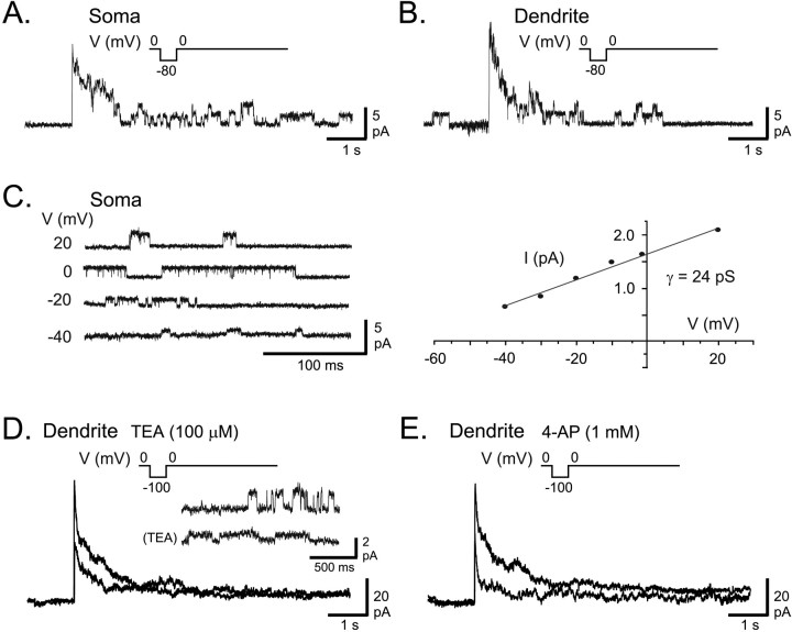
Fig. 1.
AptKv3.3 channels in ELL pyramidal cells.A, B, Outside-out patch recordings of K+ channels isolated from a pyramidal cell soma (A) and an apical dendrite 150 μm from the soma (B) in the presence of normal extracellular aCSF. A 7 sec depolarizing step from −80 to 0 mV produces a fast activation of K+ channels that subsequently inactivate over 1–2 sec to reveal a unitary conductance level for channels in the patch. C, An outside-out patch recording obtained from somatic membrane and stepped from a holding potential of −90 mV to the indicated potentials reveals K+ channels with a conductance of 24 pS. D, E, A macropatch K+ current in an outside-out recording isolated from a pyramidal cell apical dendrite (100 μm from soma) evoked by a depolarizing step from −100 to 0 mV for 7 sec. Outward current was blocked by 100 μm TEA (D).Inset shows single-channel recordings from another dendritic outside-out patch recording at 0 mV (100 μm from soma) with a substantial reduction of single-channel conductance by 100 μm TEA. E, After washout of TEA, outward current from the same dendritic patch in D is blocked by perfusion of 1 mm 4-AP. Currents in A,B, D, and E were leak-subtracted, and capacitance artifacts were removed by digital subtraction.

