
Fig. 3.
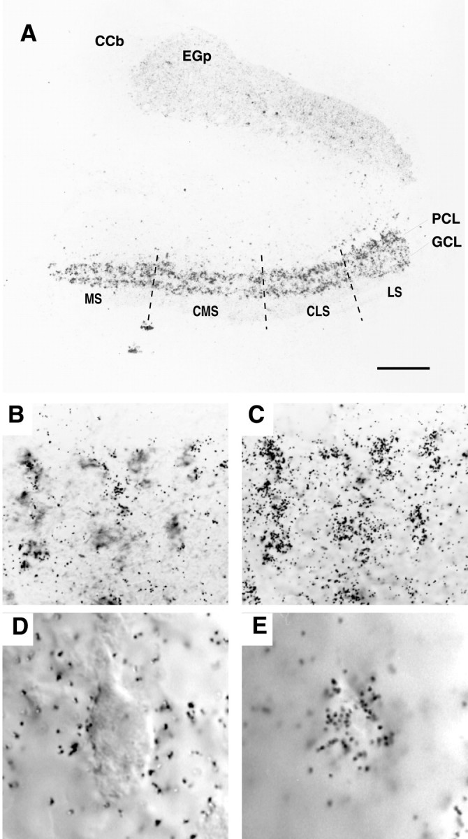
The expression of AptKv3.3 mRNA in the hindbrain. Tissue sections from hindbrain were hybridized with AptKv3.3 RNA probe. The distribution of silver grains over pyramidal and granule cell layers is indicated in A. B andC show that AptKv3.3 is expressed in all of the pyramidal cells. D and E illustrate localization of silver grains over the pyramidal cell somata.A, In situ hybridization of AptKv3.3 mRNA in the hindbrain at low power demonstrates prominent expression in the ELL and lighter expression in the adjacent caudal lobe of the cerebellum (eminentia granularis posterior; EGp) and the corpus cerebelli (CCb). The four topographic maps of the ELL are indicated: MS, medial segment;CMS, centromedial segment; CLS, centrolateral segment; LS, lateral segment. Label is dense over the entire extent of the ELL pyramidal cell (PCL) and granule cell (GCL) layers. Scale bar, 200 μm. B, A section of the pyramidal cell layer showing a cluster of pyramidal cell somata viewed under DIC optics. Scale bar, 25 μm. C, The position of silver grains in the micrograph shown in B when viewed at the plane of emulsion illustrates dense labeling positioned over individual pyramidal cell somata. D, A pyramidal cell viewed under DIC optics at higher magnification. Scale bar, 10 μm.E, Corresponding image as that shown in Dviewed at the plane of emulsion illustrates the restriction of grains to the somatic region of a pyramidal cell.