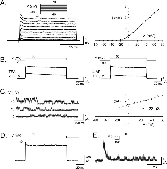
Fig. 5.
Expression of AptKv3.3 currents in HEK tsA201 cells. A, Whole-cell recording of currents expressed 1 d after AptKv3.3 cDNA transfection reveals an outward rectifying K+ current for command steps from −90 to 70 mV (10 mV steps after a 10 msec prepulse to −90 mV). AptKv3.3 K+ current exhibits a fast activation that reaches a characteristic early peak of ∼5 msec duration that subsequently relaxes to a steady-state current and fast deactivation on stepping back to −60 mV. Little steady-state inactivation is apparent after 60 msec. The current–voltage relationship indicates outward rectification, a high threshold for initial activation of −10 mV, and little saturation for steps up to 70 mV. B, Whole-cell AptKv3.3 currents are highly sensitive to micromole concentrations of externally applied TEA and 4-AP. C, An outside-out patch recording of AptKv3.3 single channels stepped to different steady-state potentials reveals a single channel slope conductance of 23 pS. D, An outside-out macropatch recording of AptKv3.3 current reveals a very similar response as found for whole-cell currents >100 msec (compare with A).E, An outside-out patch recording of AptKv3.3 single channels indicates a fast and maximal activation of channels over the initial 200 msec followed by inactivation during a 7 sec depolarizing command step from −100 to 0 mV. Capacitance artifacts inD and E were digitally subtracted.

