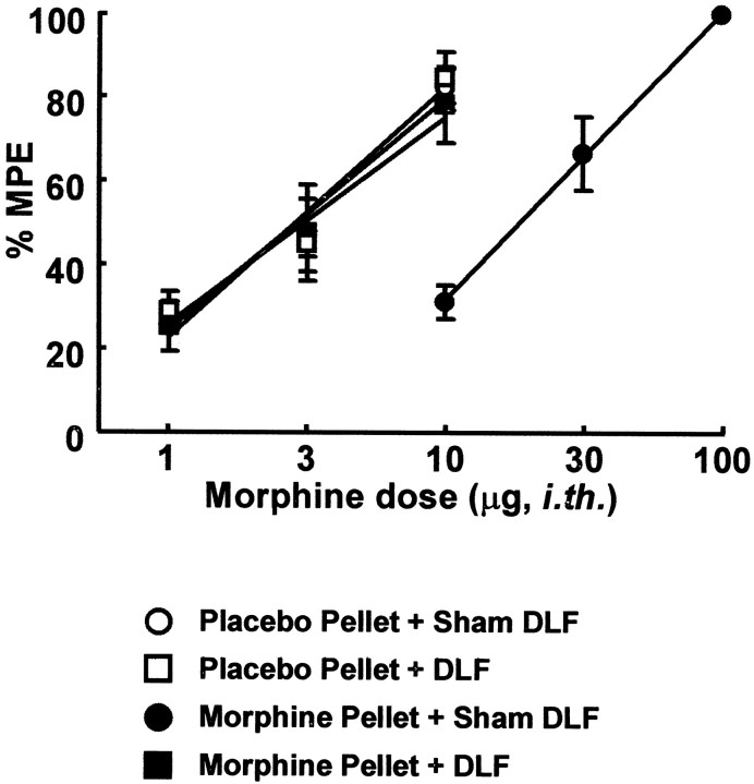
Fig. 6.
Male Sprague Dawley rats received either bilateral lesions of the DLF or sham lesions. In addition, 2 d after surgery, animals received either subcutaneous implantation of placebo pellets or morphine (75 mg) pellets. After 7 d of pellet exposure, antinociceptive dose–response functions for intrathecal morphine were generated in the 52°C water tail flick test at the time of peak effect of morphine (30 min). The following groups were used: placebo-pelleted rats with sham DLF lesions (○), placebo-pelleted rats with DLF lesions (■), morphine-pelleted rats with sham DLF lesions (●), and morphine-pelleted rats with DLF lesions (▪). The dose–effect curve for intrathecal morphine in the morphine-pelleted group was shifted significantly to the right of that for the placebo-pelleted group. This dose–effect curve of the morphine-pelleted group with DLF lesions was not different from that of the placebo-pelleted groups.

