
Fig. 1.
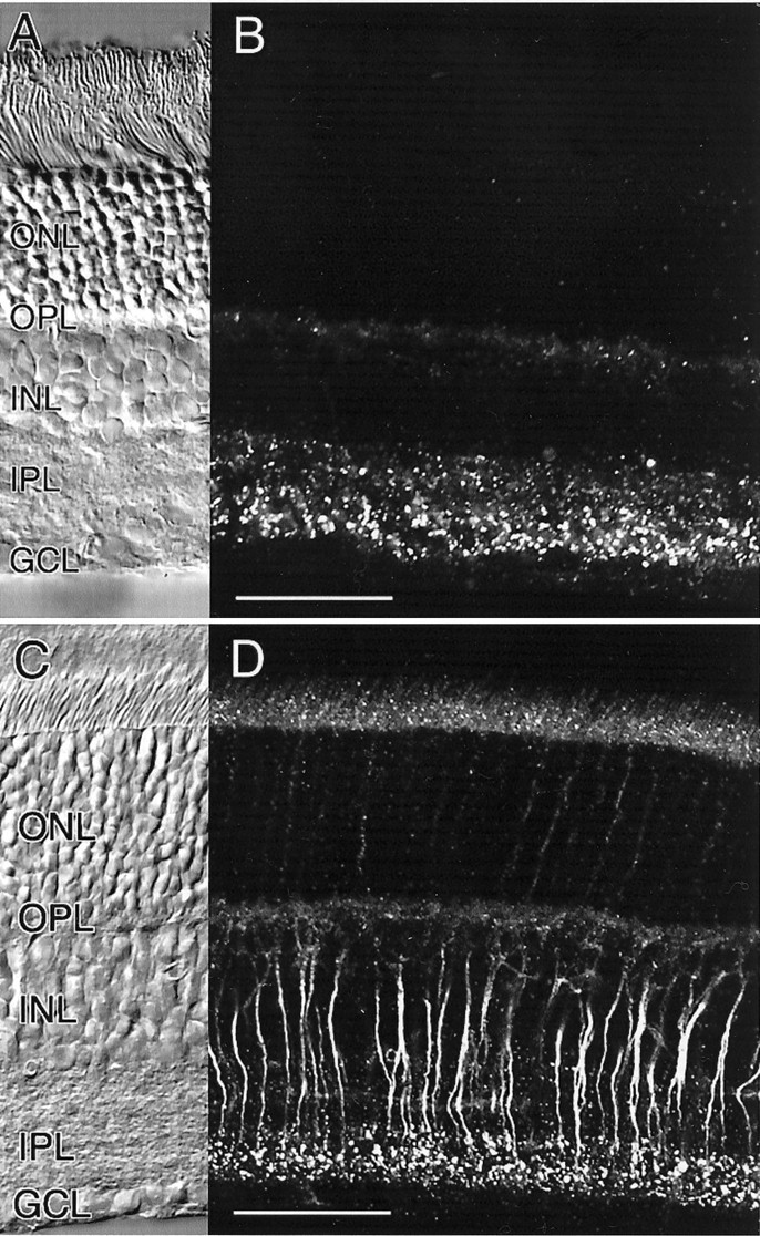
Immunocytochemical localization of Cx36 in the retina of rat and mouse. A, B, Pattern of Cx36 immunoreactivity in vertical sections of the rat retina. The corresponding Nomarski image is shown in A.C, D, Pattern of Cx36 immunoreactivity in vertical sections of the mouse retina. The cytoplasmic staining of rod bipolar cell axons is attributable to paraformaldehyde fixation (see Materials and Methods). The corresponding Nomarski image is shown inC. Scale bars: A–D, 50 μm.