
Fig. 5.
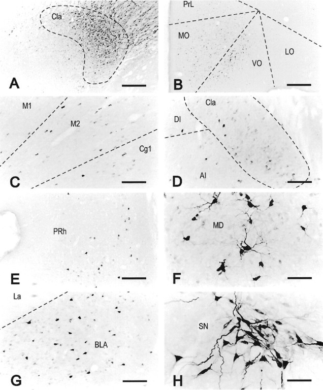
Localization of FG immunoreactivity in rat 99143. Representative bright-field microphotographs of coronal sections showing an ejection site that occupied the major part of the anterior claustrum (A) and the distribution of FG-immunoreactive neurons in the orbital cortex (B), motor and cingulate cortices (C), middle part of the claustrum (D), perirhinal cortex (E), central part of the mediodorsal thalamic nucleus (F), basolateral amygdaloid nucleus (G), and substantia nigra (H). The orientation of all images is such that the left side of the image is to the lateral side of the brain, right side to medial, and top side to dorsal. All other abbreviations are as indicated in previous figures. SN, Substantia nigra. Scale bars:A, B, 435 μm;C–E, G, 174 μm;F, H, 87 μm.