
Fig. 6.
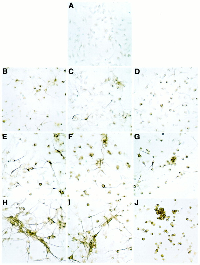
Staining of PDGF-AA-treated cortical stem cells with neuronal markers. After fixation, the cells were stained with MAP2 antibodies. Control cells, grown in the presence of FGF2, were fixed at the start of the experiment (A). Cells were grown for 2 d (B–D), 4 d (E–G), or 6 d (H–J) without FGF2 in the absence or presence of PDGF. Parallel cultures were untreated (B, E, H), received a single dose of PDGF-AA (C, F,I), or PDGF-AA was added daily (D,G, J). The cells were photographed at 200× magnification.