
Fig. 1.
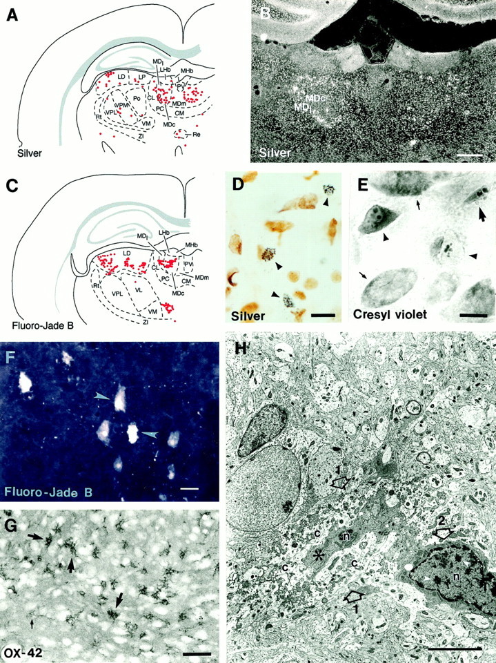
A, Computer-generated plot demonstrating the distribution of silver-positive cells in the different nuclei of the thalamus. Each red dotrepresents one silver-positive cell. B, A dark-field photomicrograph demonstrating the silver-positive cells (appear aswhite dots) in the thalamus in a rat that experienced StE 48 hr earlier (case StE10). Note the large number of silver-positive cells in the periphery of the central segment and also in the dorsal aspect of the lateral segment. C,Computer-generated plot demonstrating the distribution of Fluoro-Jade B-positive cells in the different nuclei of the thalamus. Eachred dot represents one labeled cell (case StE21). Note the similarity in the distribution with silver-positive cells in A. D, Color photomicrograph showing the appearance of silver-positive cells under bright-field illumination (arrowheads; case StE 10). Note the granular accumulation of silver deposits and shrunken appearance of the remains of the cell. Most of the undamaged cells are glial cells. E, High-power bright-field photomicrograph of neurons in the posterior pole of the anteromedial nucleus in cresyl violet staining (StE11). Note the fragmented appearance of nuclei in cells with pyknotic (arrowhead) or shrunken (large arrow) somata. Under fluorescence light (FITC filters) these same neurons had a pale appearance and could be easily distinguished from surrounding neurons (data not shown).F, A confocal image showing the appearance of Fluoro-Jade B-positive cells in the mediodorsal nucleus (arrowheads; case StE21). G,Ameboid-shaped activated microglia (thick arrows) in the central segment of the mediodorsal nucleus in preparations stained with an antibody raised against OX-42. Thin arrow points to an inactive OX-42-positive microglia. H, An electron micrograph showing a cell body (1 with open arrows) undergoing lysis. In the center, there is a small part of the nucleus (n) and condensed cytoplasm (asterisk), which are surrounded by lysed cytoplasm (c). Note the disintegration of cellular components, which is a sign of irreversible (necrotic) neuronal damage. Adjacent to the lysed cell on the right(2 with open arrow) there is a microglial cell with an irregular shape, dense cytoplasm, and clumped chromatin in the nucleus (n, white arrowheads) and on the nuclear membrane. CL, Centrolateral nucleus; CM, central medial nucleus; LD, laterodorsal nucleus;LHb, latreal habenula; LP, lateral posterior nucleus; MDc, central segment of the mediodorsal nucleus; MDl, lateral segment of the mediodorsal nucleus; MDm, medial segment of the mediodorsal nucleus; MHb, medial habenula;PC, paracentral nucleus; Po, posterior thalamic nuclear group; PV, paraventricular nucleus;Re, reuniens nucleus; Rt, reticular nucleus; VL, ventrolateral nucleus; VM, ventromedial nucleus; VPL, ventral posterolateral nucleus; VPM, ventral posteromedial nucleus;ZI, zona incerta. Scale bars: B, 500 μm;D–F, 10 μm; G, 50 μm;H, 5 μm.