Fig. 2.
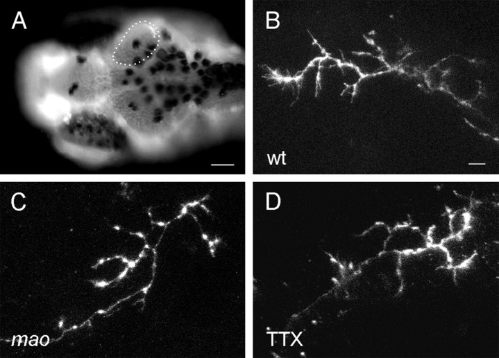
Morphology of individual RGC axons is not altered in mao mutant or TTX-treated larval tecta at 6 dpf.A, DAPI-stained tectal neuropil of a wild-type larva with a single axon terminal labeled with DiI in the posterior medial quadrant. Branching patterns of single RGC axons in wild-type (B), mao mutants (C), and TTX-treated larvae (D) visualized by DiI injection into the retina. Scale bar: A, 50 μm; B–D, 10 μm.

