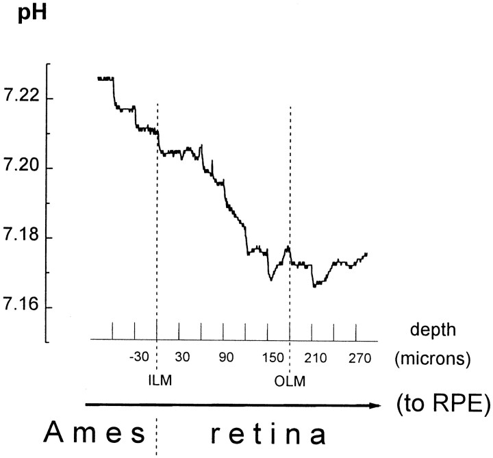
Fig. 3.
Retinal pHo varies with retinal depth. Depth profiles of retinal pHo were obtained with pH-selective microelectrodes that were advanced through the superfusate (Ames medium) and retina to the retinal pigment epithelium (toRPE) in 30 μm steps every 30 sec. Retinal pHo was lowest in the vicinity of the outer limiting membrane (OLM). The vitreal surface of the retina was defined as a position of 0. The twovertical dashed lines indicate the depth at which the microelectrode penetrated the inner limiting membrane (ILM) and OLM. pH values are shown on the y-axis. Because the retina is a metabolically active tissue that produces acid, there is a pH gradient in its vicinity. When the pH-selective microelectrode was at a distance of 600 μm or more from the retina, the recorded pHo was 7.80 (Fig. 1). When the pH electrode was closer to the retinal surface, as shown in Figure 3, the pHo recorded in the Ames medium was < 7.80.

