Fig. 5.

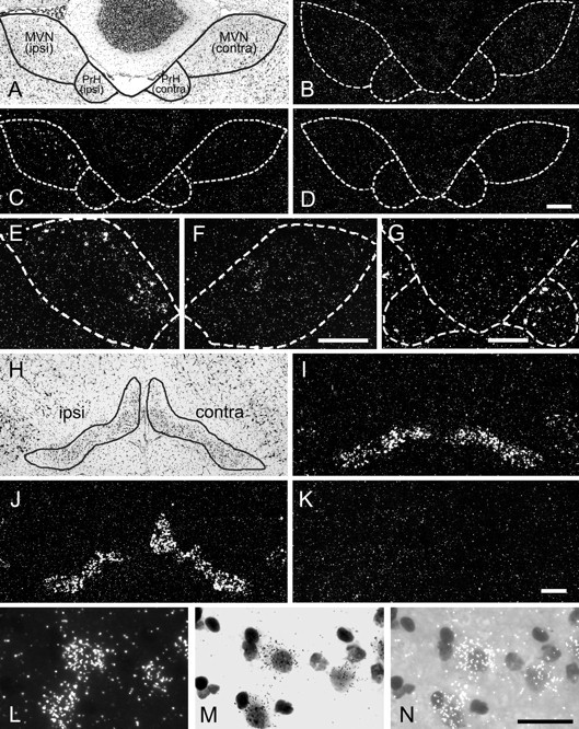
Distribution of BDNF mRNA-positive cells in the brainstem after UL. A–G, BDNF mRNA distribution in the MVN and PrH. A, A Nissl-stained section of a rat brain 6 hr after UL, indicating the boundaries of the MVN and PrH. Theleft side of the panel represents the side ipsilateral to the lesion, and the right side indicates the contralateral side. B, A section of a control rat brain hybridized with the antisense BDNF probe. Only a few BDNF mRNA-positive cells were observed in the MVN and PrH for both sides. The boundary of the nuclei was defined in an adjacent Nissl-stained section and is overlaid on B. C, A section of the rat brain 6 hr after UL, hybridized with the antisense BDNF probe. This section is adjacent to the Nissl-stained section shown inA. The boundary traced in A is overlaid on C. Many BDNF mRNA-positive cells were observed in the ipsilateral MVN and the contralateral PrH. D, A control section of the rat brain 6 hr after UL, hybridized with the sense BDNF probe. No specific signal was observed. E,F, BDNF mRNA-positive cells in the rostral part of the MVN. Shown are the ipsilateral (E) and contralateral (F) MVN taken from the section 120 μm rostral to the section shown in C. BDNF mRNA-positive cells were most prominent in the rostral part of the ipsilateral MVN. G, BDNF mRNA-positive cells in the caudal part of the PrH. The section, 160 μm caudal to the section inC, showed BDNF mRNA-positive cells in the contralateral PrH. The BDNF expression in the PrH was most obvious in the contralateral side at the caudal level. H–K, BDNF mRNA distribution in the inferior olivary complex. H, A Nissl-stained section of a rat brain 6 hr after UL, indicating the boundary of the inferior olivary complex. The boundary includes several subdivisions of the inferior olivary complex, although only the medial region of this complex was analyzed in the RT-PCR quantification experiments. I, A section of a control rat brain hybridized with the antisense BDNF probe. BDNF mRNA-positive cells were observed in the lateral region rather than the medial region of the inferior olivary complex. J, A section of the rat brain 6 hr after UL, hybridized with the antisense BDNF probe. The section is adjacent to the Nissl-stained section shown in H. BDNF mRNA-positive cells were observed in the medial region of the complex in the contralateral side as well as in the lateral region in both sides. K, A control section of the rat brain 6 hr after UL, hybridized with the sense BDNF probe. L–N, Higher magnification of BDNF mRNA-positive cells in the inferior olivary complex. The cells in the contralateral side of the medial region of the inferior olive are enlarged and shown in dark field (L), bright field (M), and bright field with epi-illumination (N). Silver grains were concentrated around lightly Nissl-stained neuronal nuclei. Scale bars:A–D, 100 μm; E, F, 100 μm; G, 100 μm; H–K, 100 μm;L–N, 50 μm.
