
Fig. 2.
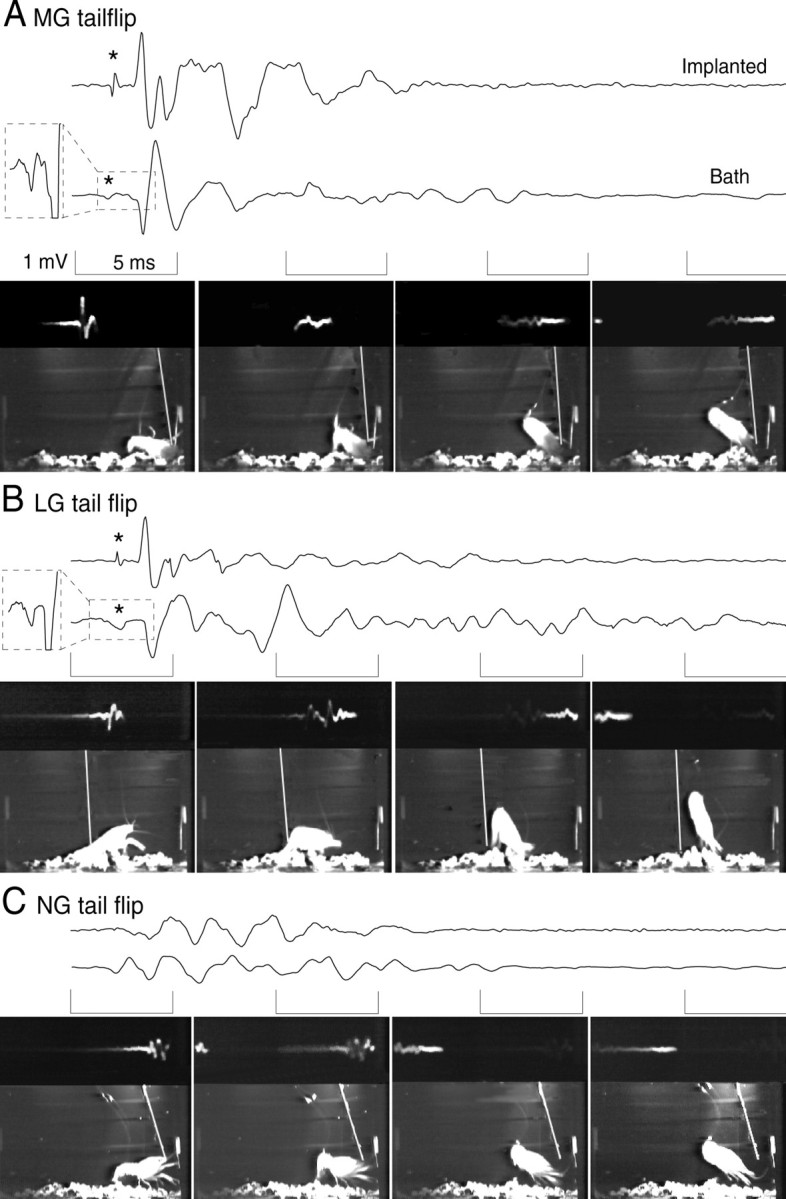
Digitized recordings from the implanted (top traces) and bath (bottom traces) electrodes with simultaneously recorded video frames of the tail-flip behavior of the animal. Each video framealso displays the reflected oscilloscope trace of the bath recording (at the top of each frame; the frames are each left-right reversed so that increasing time of the oscilloscope trace is fromleft to right), the animal, and the stimulus probe (white diagonal line). Thebracketed periods of each tracecorrespond to the period of the frame displayedbelow. A, MG tail-flip response caused by a phasic probe stimulus to the front of the animal. The field potential includes the MG giant spike potential (* and magnified in thedashed box inset to the left of the trace), the large, phasic MoG potential, and the lower-amplitude potentials produced by the FF motor neurons and FF muscles. B, LG tail-flip response caused by a phasic probe stimulus to the abdomen. As for MG, the LG field potential includes the LG spike potential (* and magnified in the dashed box inset), the MoG potential, and the FF motor neuron and FF muscle responses. C, NG tail-flip response to a nonphasic probe stimulus to the thorax. The field potential consists only of FF motor neuron and FF muscle potentials. No giant spike potential was recorded.