
Fig. 5.
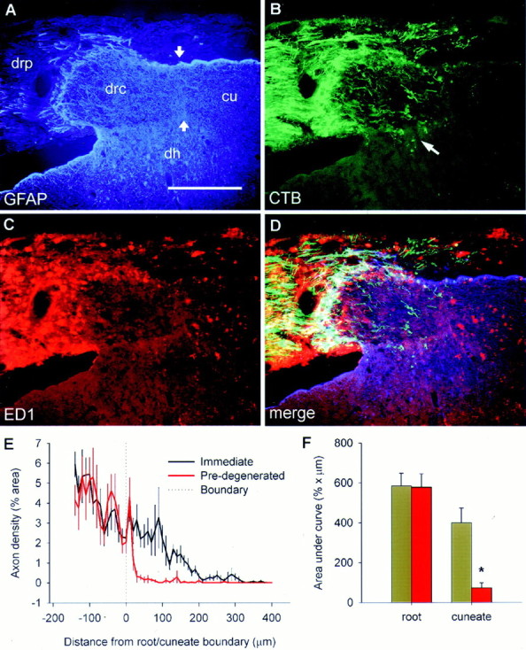
The state of degeneration dictates regenerative success. Images are from a single section taken from a rat that had received a dorsal root resection of C4–C6 and C8–C2, sparing C7 for 1 week. The C7 root was cut and allowed to reanastomose 1 week after the initial surgery, an NT-3 pump was implanted, and the rat survived for a further week. A, GFAP immunohistochemistry, showing the relationship between the peripheral and central parts of the dorsal root (drp, drc), the dorsal horn (dh), and cuneate fasciculus (cu). B,CTB-labeled axon ingrowth occurs as it does after immediate NT-3 treatment within the central part of the root (which has been degenerating for 1 week), but fails to penetrate the degenerating cuneate fasciculus (which has been degenerating for 2 weeks).Arrow indicates axon growth into the dorsal horn.C, ED-1 immunohistochemistry showing heavy invasion of the peripheral nerve and cuneate fasciculus by phagocytic cells, but with a lack of ED-1 staining in the central part of the root into which axons have regenerated. D, Merged images.E, Mean ± SEM axon density either side of the border between the central part of the dorsal root and the cuneate fasciculus (A, arrows). F, Area under the curves in D showing a significant failure of axons to penetrate the degenerating cuneate fasciculus. Scale bar, 300 μm.