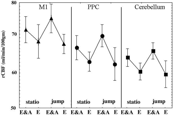
Fig. 6.
rCBF mean values (SD vertical bars), in each experimental condition, for the three cerebral regions showing significant activation in the hand error correction contrast (E&A, eye and arm pointing; E, eye alone;statio, stationary trial; jump, jump trial). These regions are the primary motor cortex (black triangles, left curve; Talairach coordinates: −30, −26, 57), the posterior parietal cortex (black circles, middle curve; −41, −44, 58), and the anterior parasagittal cerebellar cortex (black squares, right curve; 11, −45, −20).

