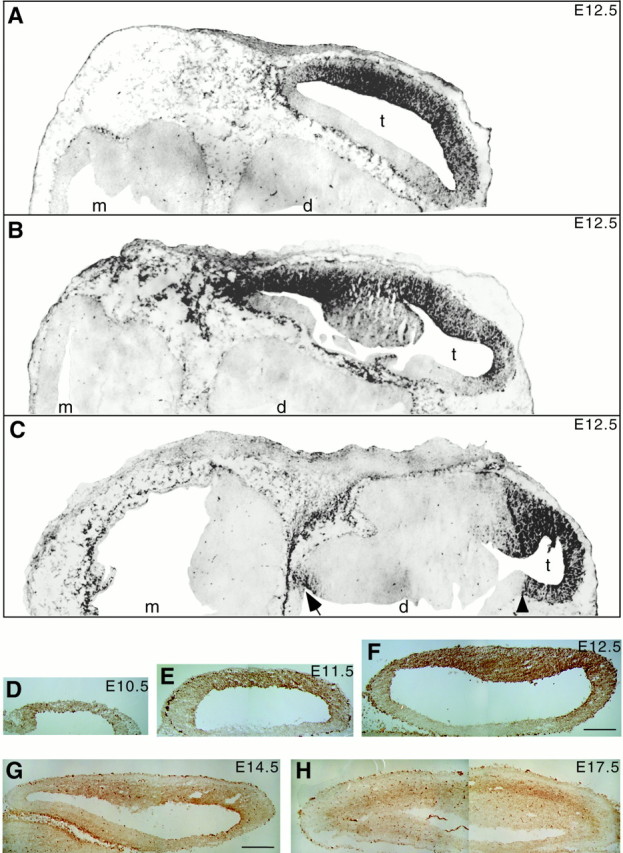
Fig. 1.

25H11 immunoreactivity in the embryonic mouse brain. Immunoperoxidase staining (A–C,black; D–H, brown) using mAb 25H11 on cryosections of embryonic mouse brain.A–C, E12.5, transverse sections at dorsal (A), intermediate (B), and ventral (C) level; to allow presentation at higher magnification (same for A–C), only one half of each section is shown. Note the immunostaining in the telencephalon (t), which is much stronger laterally than medially; an apparent boundary of immunostaining in the medial telencephalon is indicated by the arrowhead inC. Diencephalon (d) and mesencephalon (m) lack 25H11 immunostaining, except for a region that will give rise to the hypothalamus (C, arrow). Outside the brain, mesenchymal cells are immunoreactive. Staining of the choroid plexus and of scattered cells in the diencephalon and mesencephalon was also seen in sections carried through the peroxidase reaction without previous incubation with primary and secondary antibodies (data not shown), and hence reflects endogenous peroxidase activity. D–H, Transverse sections of telencephalic vesicles at E10.5 (D), E11.5 (E), E12.5 (F), E14.5 (G), and E17.5 (H). Anterior is to the right of each panel. The strongly stained cells contain endogenous peroxidase activity (see also Fig.6F). Magnifications are the same forD–F and for G and H.Scale bars: F, 100 μm; G, 200 μm.
