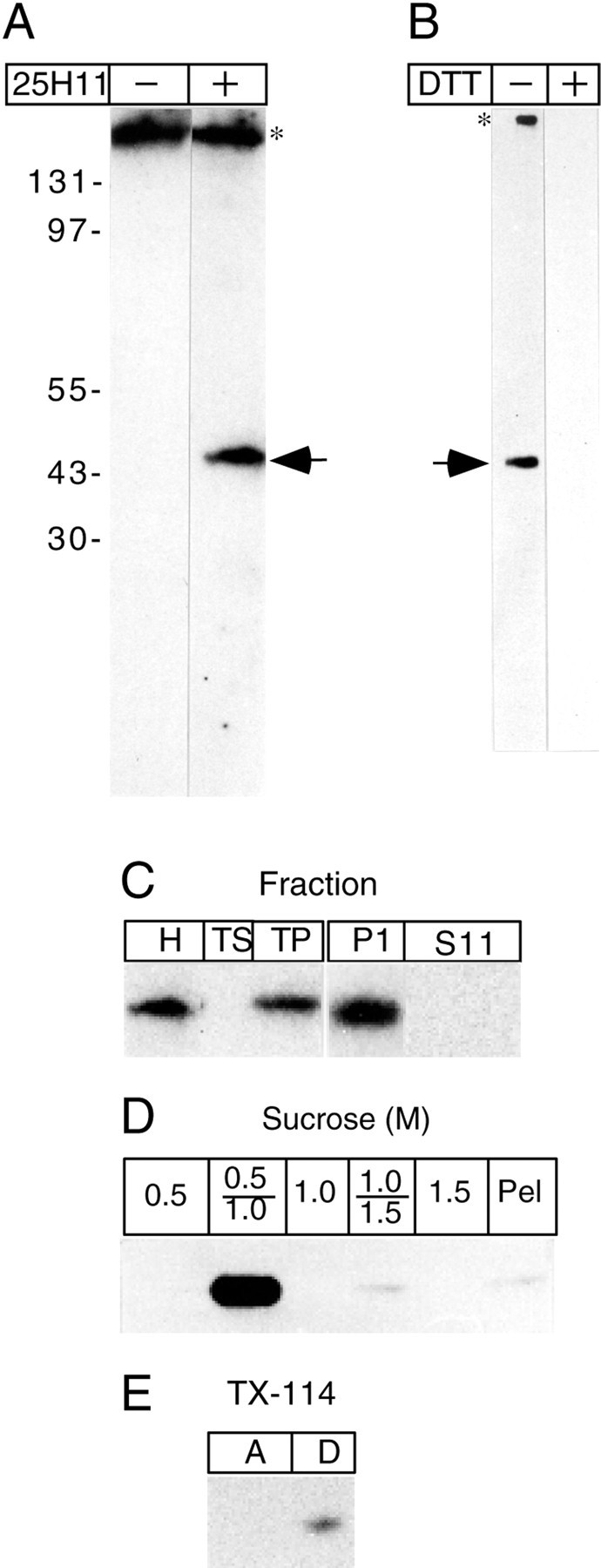
Fig. 2.

The 25H11 antigen is a 47 kDa integral membrane protein. A, Immunoblot after nonreducing SDS-PAGE of the total homogenate of ≈1.7 mouse E12.5 telencephali without (−) and with (+) 25H11 antibody followed by [125I]Protein A. B, After nonreducing SDS-PAGE of the total homogenate of ≈0.7 mouse E13.5 telencephali, nitrocellulose filters were treated without (−) and with (+) 10 mm DTT at 55°C, followed by immunolabeling with 25H11 antibody and the ECL detection system.A, B, Arrows, 25H11 antigen; asterisks, endogenous protein (presumably mouse Ig) recognized by the secondary antibody. The positions of molecular weight markers are indicated inA. C, 25H11 Immunoblots of various fractions prepared from the homogenate of ≈3 E12–E13 mouse telencephali. Left immunoblot,H, homogenate; TS, total supernatant; TP, total pellet. Right immunoblot, P11 and S11, pellet and supernatant, respectively, obtained from the total pellet by pH 11 carbonate treatment. D, 25H11 immunoblot of fractions from a floatation sucrose step gradient of pH 11 carbonate-treated membranes prepared from seven E13.5 mouse telencephali. The entire 0.5, 1.0, and 1.5 m sucrose phases and the two interfaces were analyzed. Pel, Pellet. E, 25H11 immunoblot of the aqueous (lane A) and detergent (lane D) phase obtained after phase condensation of a Triton X-114 extract of pH 11 carbonate-treated membranes prepared from ≈1 E13.5 mouse telencephalon.
