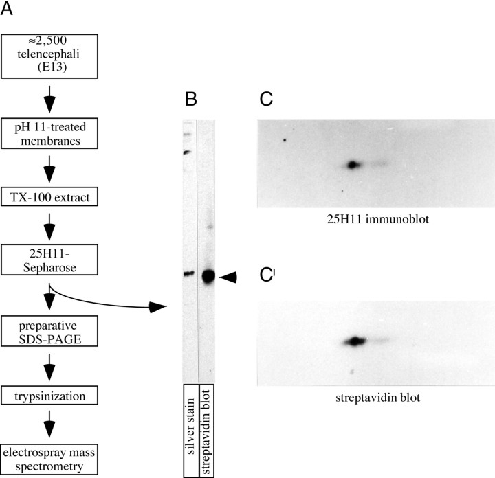
Fig. 3.
Purification of the 25H11 antigen.A, Flow scheme of the purification of the 25H11 antigen from E13.5 mouse telencephali and of its further processing for nanosequencing. For details, see Materials and Methods.B, Aliquots (0.2%) of the eluate from the 25H11-Sepharose (A) were analyzed by SDS-PAGE followed by silver staining or were biotinylated and analyzed by SDS-PAGE followed by streptavidin blotting. Arrow, 25H11 antigen. C, C′, The 25H11 antigen was purified through the 25H11-Sepharose step as described inA, except that only ≈140 E13.5 mouse telencephali were used. An aliquot (25%) of the eluate from the 25H11-Sepharose was PEG-precipitated and biotinylated, and an aliquot of it (80%) was analyzed by two-dimensional PAGE (acidic, right) followed by immunoblotting with the 25H11 antibody (C, ≈5 min time ECL exposure). The nitrocellulose was incubated in reducing condition to remove the antibody and reprobed with streptavidin-HRP (C′, ≈1 min time ECL exposure).

