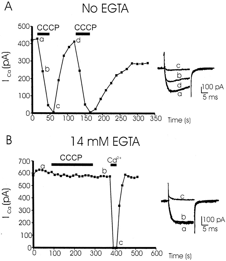
Fig. 1.
CCCP provokes the inhibition ofICa in cells dialyzed with an EGTA-free intracellular solution, but not in the presence of EGTA. The two cells of A and B were voltage clamped at −80 mV and dialyzed with 14 mm EGTA (B) or with an intracellular solution without EGTA (A). To generate inward whole-cell Ca2+ channel currents (10 mm Ca2+ in the extracellular solution), cells were stimulated with 20 msec test depolarizing pulses to +10 mV at 15 sec intervals. Each black square shows the amplitude of peak ICa (ordinates) as a function of the time course shown in the abscissa. CCCP (2 μm) and Cd2+ (100 μm) were added with the extracellular solution (that continuously superfused the cells) during the time periods indicated by the horizontal black bars. Insets at the right in A and Bshow original current traces taken at the times shown by small letters. These are original records of two typical experiments, of eight for each protocol (see Results for averaged data).

