
Fig. 10.
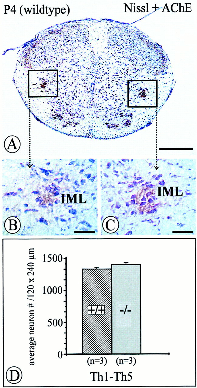
Quantification of acetylcholinesterase-positive, Nissl-stained IML neurons within an area of 120 × 240 μm of the intermediate gray in the spinal cord at P4. Numbers of IML neurons inNT-4-deficient mice (C, D) were not significantly reduced compared with controls (B, D) at this age. Data are given as mean ± SEM. Scale bars: A, 500 μm;B, C, 50 μm.