
Fig. 8.
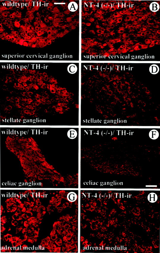
Comparison of TH-IR in the SCG (A,B), SG (C, D), CG (E, F), and adrenal medulla (G, H) ofNT-4-deficient mice and wild-type littermates. Although TH-IR is not overtly altered in the SCG, the SG, CG, and adrenal medulla reveal a qualitative reduction of TH staining intensity. Scale bars: A–D, G, H, 50 μm;E, F, 100 μm.