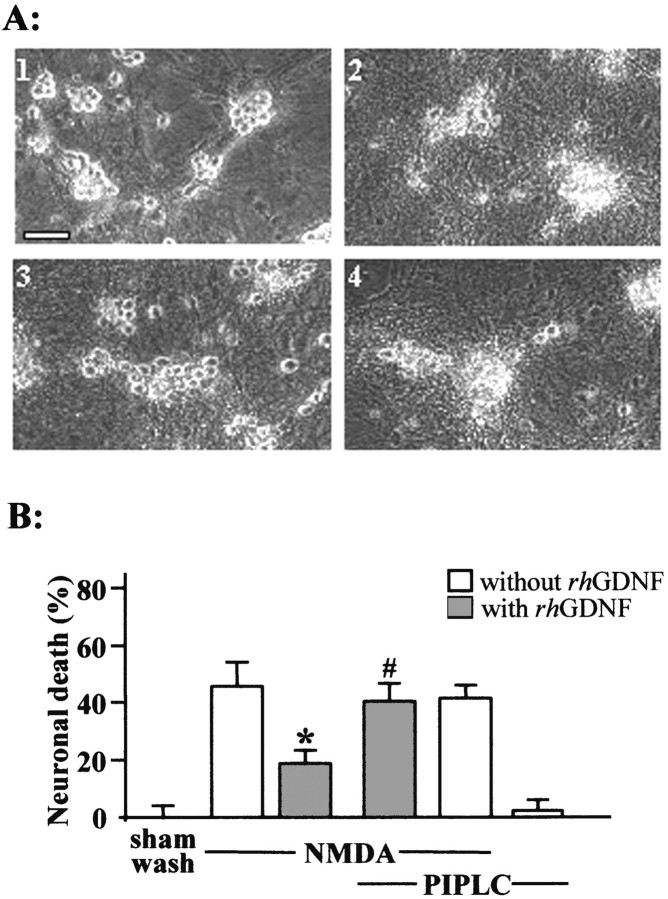
Fig. 5.
A GPI-linked protein mediates the neuroprotective activity of rhGDNF. A, Phase-contrast photomicrographs of mixed neuron–glia cortical cultures pretreated or not with PIPLC (0.3 IU/ml for 2 hr at 37°C) after NMDA application (12.5 μm) in the presence or absence of rhGDNF. Top left, Sham-washed cultures; top right, NMDA-treated cells; bottom left, NMDA-treated cells coincubated with rhGDNF (10 ng/ml); bottom right, cells pretreated with PIPLC and incubated with NMDA and rhGDNF.B, Neuronal death percentage was estimated by LDH release (mean ± SEM; n = 12) after a 24 hr exposure to NMDA (12.5 μm) in mixed neuron–glia cortical cultures in the presence (gray bars) or absence (white bars) of rhGDNF and pretreated or not pretreated with 0.3 IU/ml PIPLC for 2 hr at 37°C. * indicates significantly different from NMDA; # indicates significantly different from NMDA plus GDNF by ANOVA, followed by Bonferroni–Dunn's test (p < 0.05).

