
Fig. 8.
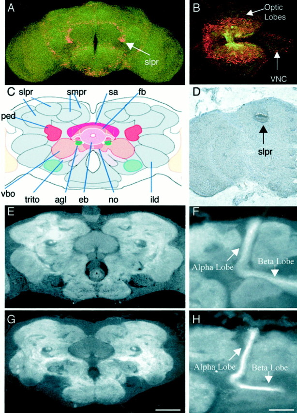
tutl is expressed in the larval and adult CNS, but no morphological defects were detected in these structures. A,B, tutl reporter gene expression in the adult brain and the larval brain represented by β-gal expression from the lacZ gene on the l(2)01085 P-element (red). The CNS was stained with mAb BP102 for reference purposes (green). Oriented dorsal out of the page and anterior up (A) or to the left (B). C, Frontal schematic of an adult Drosophila brain from the Flybrain website (www.flybrain.org). ped, Pedunculus;fb, fan-shaped body; vbo, ventral body;smpr, superior medial protocerebrum;trito, tritocerebrum; sa, superior arch,eb, ellipsoid body; no, nodulus;ild, inferior lateral deutocerebrum; igt, antennoglomerular tract; vbo, ventral body.D, DNA in situ hybridization of a frontal adult brain histology slice demonstrating confined tutlexpression in the adult brain to the region of the superior lateral protocerebrum. Note that this expression is consistent with that seen in A. E, Wild-type adult brain frontal slice stained with mAb BP102 to visualize structures labeled inC. F, Wild-type adult brain frontal slice stained with mAb BP102 to visualize the α and β lobes of the mushroom bodies. G, tutl2 adult brain frontal slice stained with mAb BP102 to visualize structures labeled inC. No morphological abnormalities were found.H, tutl2 adult brain frontal histology slice stained with mAb BP102 to visualize the α and β lobes of the mushroom bodies. No morphological abnormalities were found in any of the lobes of the mushroom bodies. Scale bars: A, 100 μm; B, 90 μm; C–E, G, 60 μm; F, H, 30 μm.