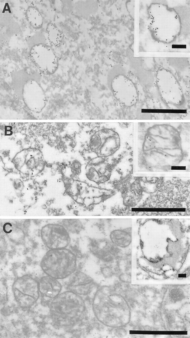
Fig. 4.

Ultrastructural examination of a pyramidal neuron in the hippocampus after in situ hybridization by using probes to wild mtDNA (wild type 1) in AD (A, B) or mtDNAΔ5kb (chimeric probe) (insets, A, B). The high density of gold particles was seen inside the vacuolar portions of lipofuscin granules, which likely represents autophagocytosis of damaged mitochondria in AD (A) and to a lesser extent in controls (C, inset). In contrast, mitochondria with cristae in both AD (B) and control cases (C) showed a lower level of mtDNA labeling (C). Scale bars: 1 μm; 0.25 μm (insets).
