
Fig. 2.
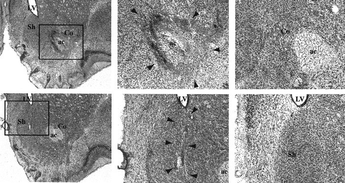
Photomicrographs showing thionin-stained coronal sections through the nucleus accumbens. Top, Representative core lesions. Bottom, Representative shell lesions. Left, Images of the general region of the NAC (40× magnification) of core-lesioned (top) and shell-lesioned (bottom) animals. Middle, Greater magnification of the region indicated by the outlined boxes in the left images; arrowheads indicate lesion boundaries.Right, High-magnification photographs of the region of the lesions shown in the middle images (in lesioned animals) but in sham-lesioned animals. Allimages are from slices taken at ∼2.6 mm anterior to bregma. ac, Anterior commissure; Co, NAC core; LV, lateral ventricle; Sh, NAC shell.